

The cardiac cycle is one of the most predictable topics in the Circulation chapter of MDCAT Biology. Questions ask how long one cycle lasts, which valves are open in ventricular systole, what makes the lub and the dub, and where the heartbeat starts. Each of these is a one-line fact, but the facts only stick when you can picture the whole beat. This guide walks you through one heartbeat in order, with the timings, the valves, the heart sounds, the conducting system and double circulation, all checked against ePrepare's notes and FSc books.
The short answer: One cardiac cycle is one contraction (systole) and one relaxation (diastole) of the heart, and it takes about 0.8 second, which gives about 75 beats per minute. The order is atrial systole (0.1 s) → ventricular systole (0.3 s) → diastole of the whole heart (0.4 s). Lub is the AV valves closing at the start of ventricular systole; dub is the semilunar valves closing at the start of diastole. Each beat starts in the SA node and spreads through the AV node, bundle of His and Purkinje fibres.
What is the cardiac cycle?
The heart fills with blood from the large veins, then contracts to push that blood into the arteries. One full round of filling and pumping is one heartbeat, called a cardiac cycle. These contractions and relaxations are cyclic and rhythmic, and they never stop.
Key definitions
- Cardiac cycle: the sequence of events in one heartbeat: one contraction and one relaxation of the heart.
- Systole: contraction (squeeze). Diastole: relaxation (dilate).
- Atrial systole / diastole: the atria contract / relax. Ventricular systole / diastole: the ventricles contract / relax.
- Used alone, "systole" and "diastole" usually mean ventricular systole and diastole.
- AV (atrioventricular) valves: between atria and ventricles: tricuspid on the right, bicuspid (mitral) on the left.
- Semilunar valves: at the exits of the ventricles: pulmonary valve (right ventricle → pulmonary trunk) and aortic valve (left ventricle → aorta).
Remember this: "1 + 3 + 4 = 8". Atrial systole 0.1 s + ventricular systole 0.3 s + diastole 0.4 s = 0.8 s. And "lub before dub, A before S": lub = AV valves close; dub = Semilunar valves close.
The phases of one heartbeat, in order
Valves are passive flaps. They open and close only because of pressure: blood moves from high pressure to low pressure, and a valve shuts whenever the pressure would push blood backward. Keep that rule in mind and every phase makes sense.
1. Atrial diastole (filling)
The atria are relaxed. Blood enters the right atrium from the body through the venae cavae, and the left atrium from the lungs through the pulmonary veins. As the atria fill, pressure rises and pushes the AV valves open, so blood starts flowing into the relaxed ventricles.
2. Atrial systole (about 0.1 s)
Both atria contract together and push the rest of their blood through the open AV valves into the ventricles. The tricuspid and bicuspid valves are open; the semilunar valves are closed.
3. Ventricular systole (about 0.3 s)
Just as the atria finish, the ventricles contract. Pressure in the ventricles rises above the atria and closes the AV valves, so blood cannot go back up. This closure makes the first heart sound, lub. The rising pressure then opens the semilunar valves, and blood is pushed into the aorta and pulmonary trunk.
4. Diastole of the whole heart (about 0.4 s)
The ventricles relax and their pressure falls. Blood in the aorta and pulmonary artery tends to flow back, and this closes the semilunar valves, making the second heart sound, dub. The AV valves open again, and the whole heart rests and fills for the next beat.
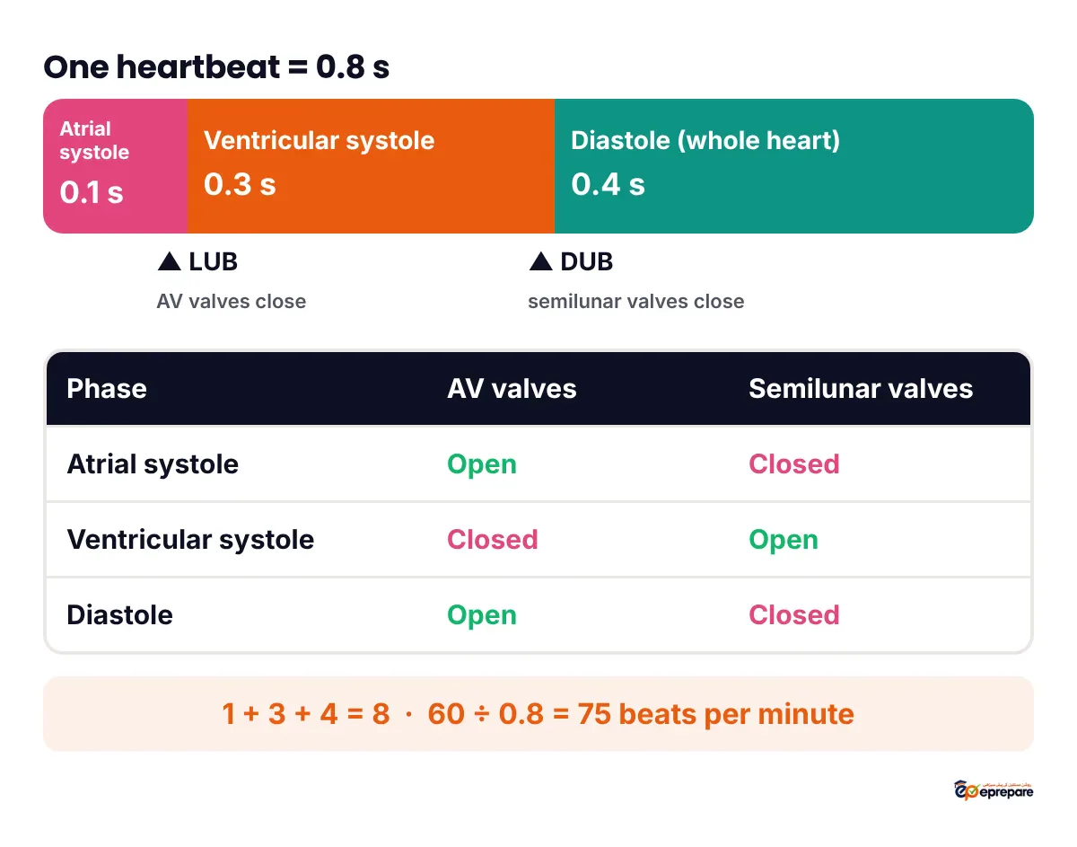
| Phase | Time | AV valves (tricuspid, bicuspid) | Semilunar valves (pulmonary, aortic) | Sound |
|---|---|---|---|---|
| Atrial systole | ~0.1 s | Open | Closed | None |
| Ventricular systole | ~0.3 s | Close at the start, stay closed | Open | Lub (AV valves closing) |
| Diastole (whole heart) | ~0.4 s | Open again | Close at the start, stay closed | Dub (semilunar valves closing) |
Timings you can work out yourself
- Atria: contract for 0.1 s, so they are relaxed for 0.8 − 0.1 = 0.7 s of each cycle.
- Ventricles: contract for 0.3 s, so they are relaxed for 0.8 − 0.3 = 0.5 s of each cycle.
- Whole heart relaxed together: about 0.4 s, half of every beat.
- Heart rate from cycle length: beats per minute = 60 ÷ cycle time. 60 ÷ 0.8 = 75 beats per minute. If one cycle took 0.6 s, the rate would be 60 ÷ 0.6 = 100; if it took 1 s, the rate would be 60.
Heart sounds: lub and dub
With a stethoscope you hear two sounds in every beat. They come from valves closing, not opening.
| Sound | Cause | When | Pitch |
|---|---|---|---|
| Lub (first sound) | Closure of the AV valves (tricuspid and bicuspid) | Start of ventricular systole | Low-pitched |
| Dub (second sound) | Closure of the semilunar valves (pulmonary and aortic) | Start of ventricular diastole | Higher-pitched |
If a valve does not close properly, some blood leaks backward and makes a different sound, called a heart murmur.
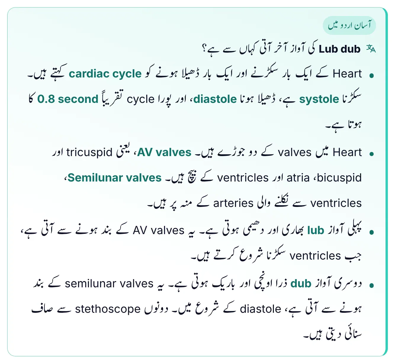
This box from ePrepare's notes asks the question students actually wonder about: where does the lub dub sound come from? Point one defines the cardiac cycle as one squeeze and one relaxation of the heart: squeezing is systole, relaxing is diastole, and a full cycle takes about 0.8 second. Point two names the two pairs of valves: the AV valves (tricuspid and bicuspid) between the atria and ventricles, and the semilunar valves at the mouths of the arteries leaving the ventricles. Point three says the first sound, lub, is heavy and low, and comes from the AV valves closing as the ventricles begin to contract. Point four says the second sound, dub, is a little higher and sharper, and comes from the semilunar valves closing at the start of diastole; both are heard clearly with a stethoscope.
The conducting system: where the beat starts
Cardiac muscle is myogenic: the beat starts inside the heart muscle itself, not in the brain. That is why a heart can keep beating for a while even after it is removed from the body. Nerves can only speed it up or slow it down. The beat is started and spread by special strands of cardiac muscle that form the conducting system.
- SA node (sinoatrial node): in the upper wall of the right atrium, near where the superior vena cava enters. Its cells make impulses faster than any other heart cells, so it sets the pace. It is the pacemaker.
- Across the atria: the impulse spreads over both atria, which contract.
- AV node (atrioventricular node): near the junction of the right atrium and right ventricle. It acts as a relay station and delays the impulse by about 0.11 s, so the atria finish emptying before the ventricles contract.
- Bundle of His (AV bundle): runs from the AV node into the interventricular septum and divides into right and left bundle branches.
- Purkinje fibres: the terminal branches, just below the endocardium of the ventricles. They are wide cardiac muscle fibres (not nerves) with many gap junctions, so they conduct very fast and make the ventricles contract together, from the apex upward.
Timings in ePrepare's notes: SA node to AV node about 0.04 s; delay inside the AV node about 0.11 s; total delay before the ventricles contract about 0.15 s. Memory trick from the notes: "Send A Huge Beat, Please" = SA node → AV node → His bundle → bundle branches → Purkinje fibres.
If the SA node fails, an artificial pacemaker, an electronic device placed under the skin, can supply impulses at a set rate. A problem in the heart's rhythm is an arrhythmia: bradycardia is a rate below 60 beats per minute, tachycardia above 100.
Double circulation: why the heart is a double pump
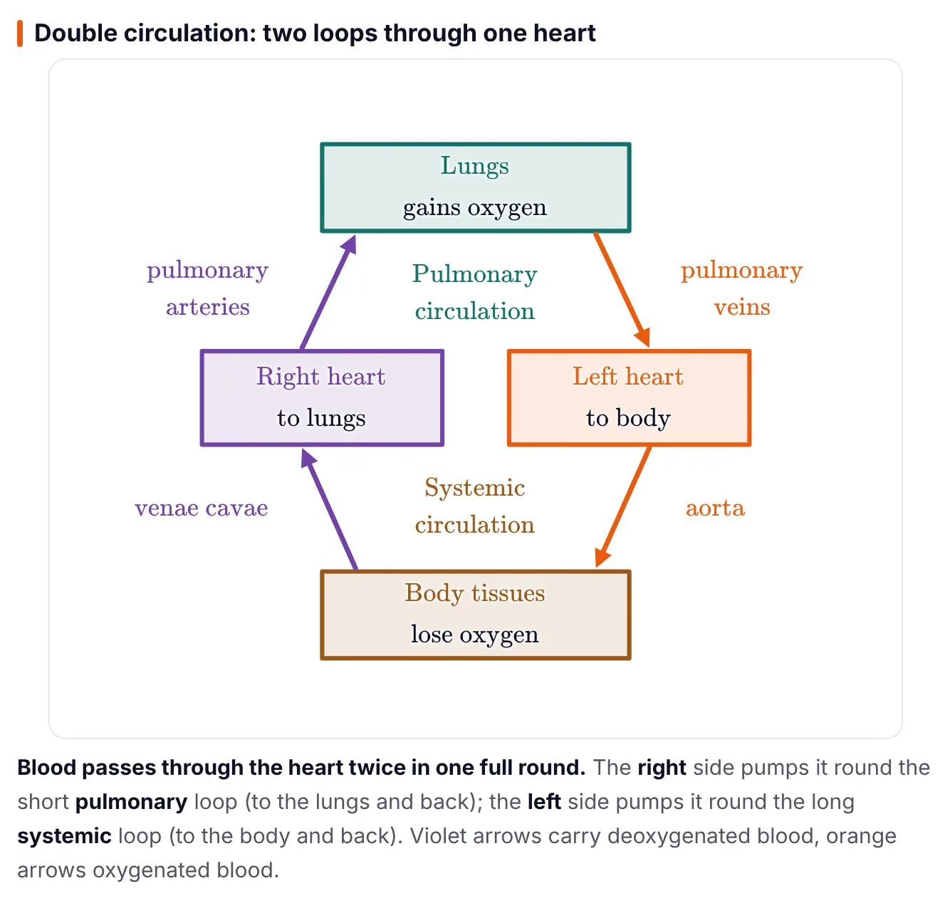
The diagram shows the heart as two pumps side by side. Start at the right heart box: it sends deoxygenated blood (violet arrow) up through the pulmonary arteries to the lungs, where it gains oxygen. Oxygenated blood (orange arrow) returns through the pulmonary veins to the left heart. That short upper loop is the pulmonary circulation. The left heart then pumps oxygenated blood through the aorta to the body tissues, which take its oxygen, and the deoxygenated blood returns through the venae cavae to the right heart. That long lower loop is the systemic circulation. In one full round, blood passes through the heart twice, which is what "double circulation" means.
The path of blood through the heart
Venae cavae → right atrium → tricuspid valve → right ventricle → pulmonary semilunar valve → pulmonary trunk → pulmonary arteries → lungs → four pulmonary veins → left atrium → bicuspid valve → left ventricle → aortic semilunar valve → aorta → body. The heart muscle gets its own blood through the coronary arteries, which drain via cardiac veins into the coronary sinus.
The pulse you feel at the wrist (radial artery, thumb side) is the pressure wave sent along the elastic arteries each time the left ventricle contracts. ePrepare's notes give the average adult pulse as about 70 to 90 beats per minute.
Question types asked in MDCAT
- Timing: length of one cycle (0.8 s), ventricular systole (0.3 s), atrial systole (0.1 s), joint diastole (0.4 s).
- Valve state: "During ventricular systole…" (AV valves closed, semilunar valves open).
- Sounds: "Lub is produced by…" (closure of AV valves). "Dub is produced by…" (closure of semilunar valves).
- Conduction: the pacemaker (SA node), its location (right atrium), the node that delays the impulse (AV node), another name for the AV bundle (bundle of His), fastest-conducting fibres (Purkinje fibres).
- Properties: "Cardiac muscle is myogenic because…" (the beat starts in the muscle itself).
- Circulation: which side sends blood to the lungs (right), which vessel carries oxygenated blood to the left atrium (pulmonary veins).
Comparison tables
| Point | Systole | Diastole |
|---|---|---|
| Meaning | Contraction | Relaxation |
| What happens | Chamber pushes blood out | Chamber fills with blood |
| Ventricles (time per cycle) | About 0.3 s | About 0.5 s |
| Atria (time per cycle) | About 0.1 s | About 0.7 s |
| Ventricular valves | AV closed, semilunar open | Semilunar closed, AV open |
| Sound at its start (ventricles) | Lub | Dub |
| Point | SA node | AV node |
|---|---|---|
| Location | Upper wall of right atrium, near the superior vena cava | Near the junction of right atrium and right ventricle |
| Job | Starts each impulse (pacemaker) | Relays the impulse and delays it |
| Key number | Fastest-firing cells in the heart | Delay of about 0.11 s |
| Passes impulse to | Atria, then AV node | Bundle of His |
Common mistakes
- "Lub is made by the semilunar valves." Lub is the AV valves closing; dub is the semilunar valves closing.
- "Heart sounds come from valves opening." They come from valves closing.
- "The AV valves are open during ventricular systole." In ventricular systole the AV valves are closed and the semilunar valves are open.
- "The heartbeat starts in the brain." Cardiac muscle is myogenic; the beat starts in the SA node.
- "The SA node is in the left atrium." It is in the upper wall of the right atrium.
- "Purkinje fibres are nerves." They are specialised cardiac muscle fibres.
- "The pulse comes from the right ventricle." It comes from the left ventricle pushing blood into the arteries.
MDCAT-style practice MCQs
1. One cardiac cycle in a normal adult at rest takes about:
- 0.1 s
- 0.4 s
- 0.8 s
- 1.8 s
Show answer
C. About 0.8 s (0.1 + 0.3 + 0.4). 0.4 s is the diastole part alone and 0.1 s is atrial systole.
2. During ventricular systole:
- AV valves are closed and semilunar valves are open
- AV valves are open and semilunar valves are closed
- Both sets of valves are open
- Both sets of valves are closed throughout
Show answer
A. Rising ventricular pressure shuts the AV valves and opens the semilunar valves, so blood goes into the aorta and pulmonary trunk.
3. The first heart sound, lub, is produced by:
- Closure of the semilunar valves
- Closure of the AV valves
- Opening of the AV valves
- Contraction of the atria
Show answer
B. Lub is the low-pitched sound of the tricuspid and bicuspid valves closing at the start of ventricular systole.
4. The second heart sound, dub, is heard at the beginning of:
- Atrial systole
- Ventricular systole
- Ventricular diastole
- Atrial diastole
Show answer
C. Dub is the semilunar valves closing when the ventricles start to relax and blood in the arteries tends to flow back.
5. If the cardiac cycle is 0.8 s long, the heart rate is:
- 60 beats per minute
- 72 beats per minute
- 75 beats per minute
- 80 beats per minute
Show answer
C. Beats per minute = 60 ÷ 0.8 = 75.
6. In one cardiac cycle of 0.8 s, the ventricles remain relaxed for about:
- 0.1 s
- 0.3 s
- 0.5 s
- 0.7 s
Show answer
C. Ventricular systole is 0.3 s, so the ventricles relax for 0.8 − 0.3 = 0.5 s. 0.7 s is the time the atria stay relaxed.
7. The impulse is delayed for about 0.11 s at the:
- SA node
- AV node
- Bundle of His
- Purkinje fibres
Show answer
B. The AV node holds the impulse so the atria finish emptying before the ventricles contract.
8. The correct order of impulse conduction in the heart is:
- AV node → SA node → bundle of His → Purkinje fibres
- SA node → bundle of His → AV node → Purkinje fibres
- SA node → AV node → bundle of His → Purkinje fibres
- Purkinje fibres → bundle of His → AV node → SA node
Show answer
C. "Send A Huge Beat, Please": SA node → AV node → His bundle → bundle branches → Purkinje fibres.
Quick revision table
| Point | Fact to remember |
|---|---|
| Cardiac cycle | One systole + one diastole; about 0.8 s; about 75 beats per minute |
| Phase timings | Atrial systole 0.1 s, ventricular systole 0.3 s, joint diastole 0.4 s |
| Relaxed time | Atria 0.7 s, ventricles 0.5 s per cycle |
| AV valves | Tricuspid (right), bicuspid/mitral (left) |
| Semilunar valves | Pulmonary (right ventricle), aortic (left ventricle) |
| Lub | AV valves close; start of ventricular systole; low-pitched |
| Dub | Semilunar valves close; start of diastole; higher-pitched |
| Murmur | Leaking valve, blood flows back |
| Conduction | SA node → AV node (0.11 s delay) → bundle of His → bundle branches → Purkinje fibres |
| Pacemaker | SA node, upper wall of right atrium |
| Double circulation | Right side → lungs (pulmonary); left side → body (systemic) |
| Rates | Bradycardia < 60, tachycardia > 100 beats per minute |
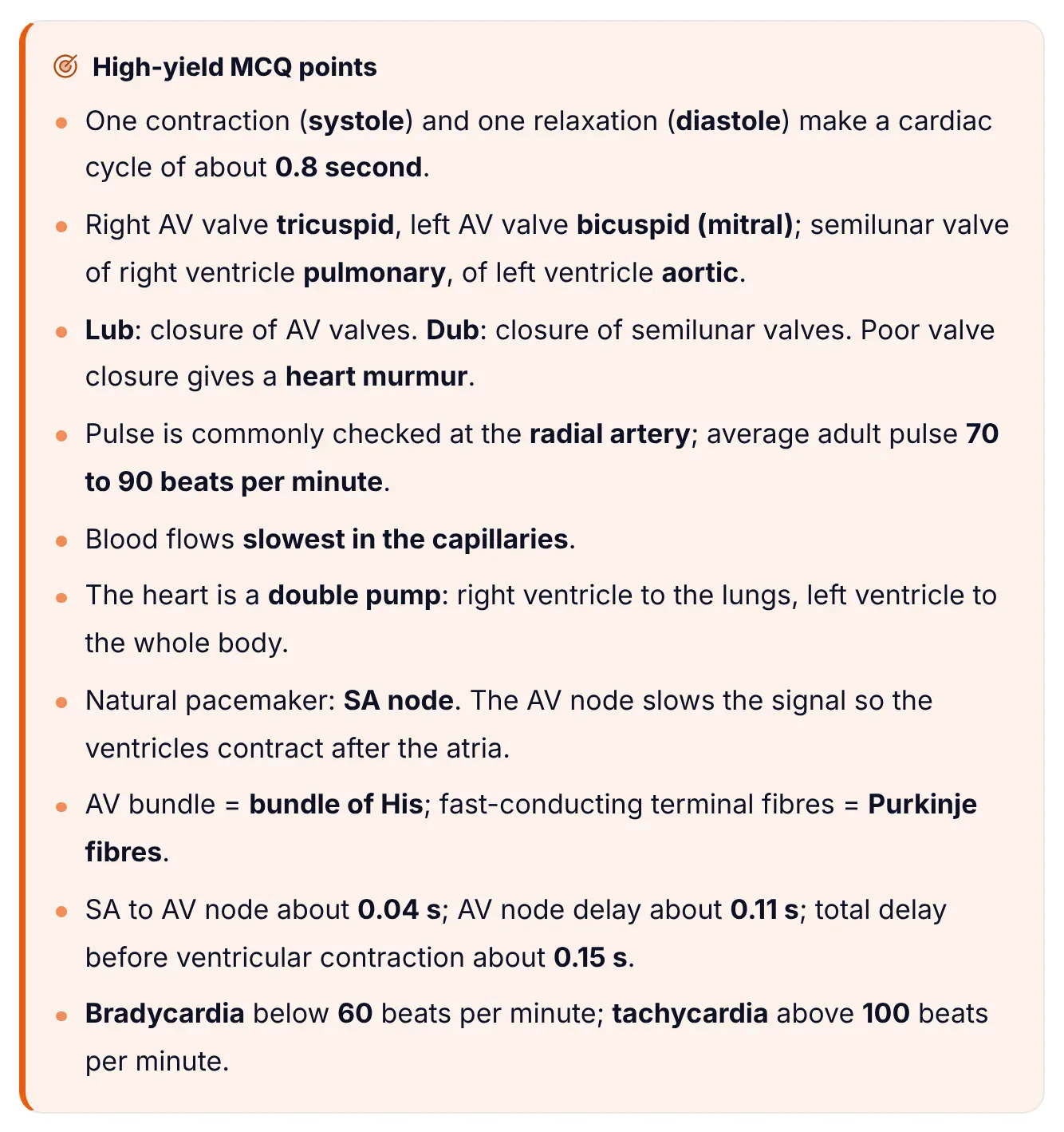
This box ends the lesson in ePrepare's notes. It packs ten facts that MDCAT turns into MCQs, from the 0.8 s cycle and the valve names to the conduction delays (0.04 s, 0.11 s, 0.15 s) and the bradycardia and tachycardia limits. The full lesson, with the cardiac cycle drawn to scale on a time line, a labelled diagram of the conducting system and 15 practice MCQs with explanations, is in ePrepare's MDCAT conceptual notes.
خلاصہ: Heart کے ایک بار سکڑنے اور ایک بار ڈھیلا ہونے کو cardiac cycle کہتے ہیں، جو تقریباً 0.8 second کا ہوتا ہے: پہلے atria سکڑتے ہیں (0.1 second)، پھر ventricles (0.3 second)، اور پھر پورا heart آرام کرتا ہے (0.4 second)۔ Lub کی آواز AV valves کے بند ہونے سے آتی ہے اور dub کی آواز semilunar valves کے بند ہونے سے۔ دھڑکن SA node سے شروع ہوتی ہے، AV node پر تھوڑا رکتی ہے، اور bundle of His اور Purkinje fibres کے ذریعے ventricles میں پھیل جاتی ہے۔
Where this fits in your preparation
Circulation is a long chapter, so revise the cardiac cycle together with heart structure and blood vessels. Our guide to high-yield MDCAT Biology chapters shows where it ranks, and the chapter-wise weightage post shows how many questions Biology carries overall. The lymphatic system links this chapter to immunity, so read our guide to the types of immunity next. Then attempt a timed chapter test in the Quiz Builder.
Frequently asked questions
How long is one cardiac cycle?
About 0.8 second in a resting adult: atrial systole about 0.1 s, ventricular systole about 0.3 s and diastole of the whole heart about 0.4 s. This works out to about 75 beats per minute.
What causes the lub and dub sounds?
Lub is the closing of the AV valves (tricuspid and bicuspid) at the start of ventricular systole. Dub is the closing of the semilunar valves (pulmonary and aortic) at the start of diastole. Both come from valves closing, not opening.
Which valves are open during ventricular systole?
The semilunar valves are open, so blood leaves the ventricles for the aorta and pulmonary trunk. The AV valves are closed, so blood cannot flow back into the atria.
Why is the SA node called the pacemaker?
Its cells make impulses faster than any other cells in the heart, so they set the rhythm for the whole heart. It lies in the upper wall of the right atrium, near the opening of the superior vena cava.
Why does the AV node delay the impulse?
The delay of about 0.11 s gives the atria time to finish emptying their blood into the ventricles before the ventricles contract.
What is double circulation?
Blood passes through the heart twice in one full round. The right side pumps it to the lungs and back (pulmonary circulation); the left side pumps it to the body and back (systemic circulation).