

Proteins do most of the work in a cell, and how a protein works depends on its shape. That is why the four levels of protein structure are a favourite MDCAT topic from the Biological Molecules chapter. Questions ask which bond holds which level, which protein has quaternary structure, and what happens when a protein is denatured. This guide explains each level simply, with the examples and traps MDCAT uses.
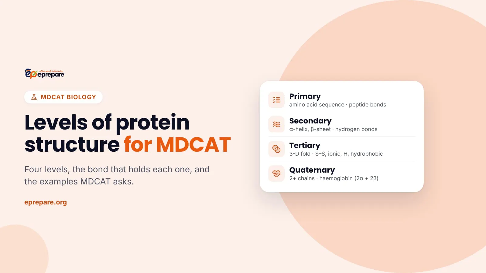
The short answer: proteins have four levels of structure. Primary is the sequence of amino acids, held by peptide bonds. Secondary is regular coiling into an α-helix or folding into a β-pleated sheet, held mainly by hydrogen bonds. Tertiary is the overall 3-D folding of one chain, held by hydrogen, ionic and disulphide bonds and hydrophobic interactions. Quaternary is two or more polypeptide chains joined together, as in haemoglobin (2 α + 2 β chains).
The building blocks: amino acids and peptide bonds
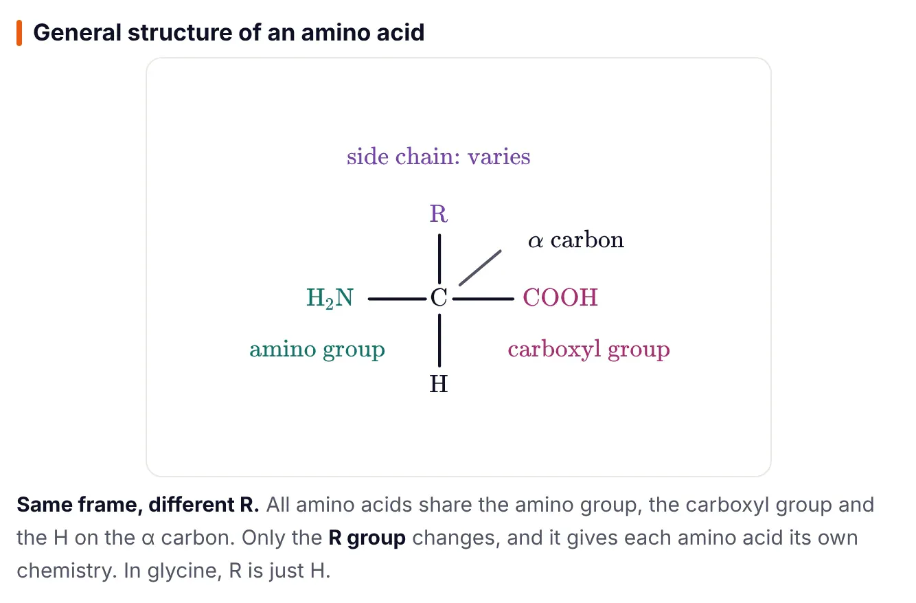
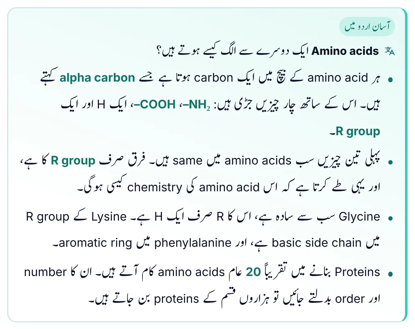
Proteins are polymers of about 20 common amino acids. Every amino acid has the same frame: a central α carbon carrying an amino group (–NH2), a carboxyl group (–COOH), a hydrogen atom and a variable side chain, the R group. The general formula is H2N–CH(R)–COOH. Only the R group changes, and it decides the chemistry of each amino acid. In glycine, R is just H.
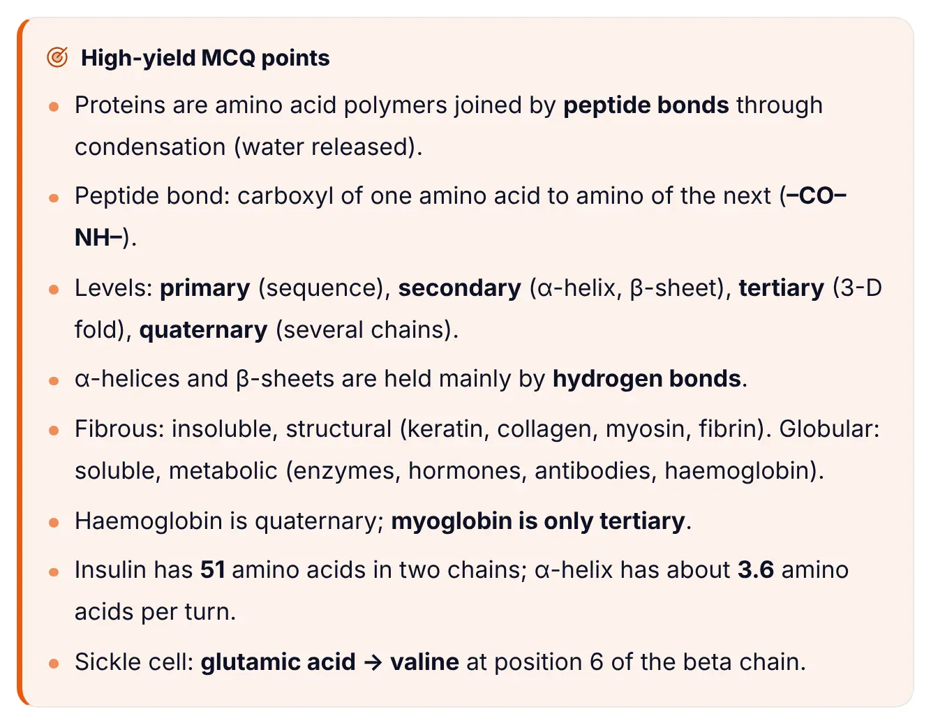
A peptide bond forms when the carboxyl group of one amino acid joins the amino group of the next by condensation, releasing a molecule of water. The bond is the –CO–NH– linkage, a C–N bond. Two amino acids make a dipeptide, three a tripeptide, and many a polypeptide. A polypeptide has a free amino end (N-terminus) and a free carboxyl end (C-terminus). Cells build polypeptides on ribosomes in the order coded by mRNA.
The four levels of protein structure
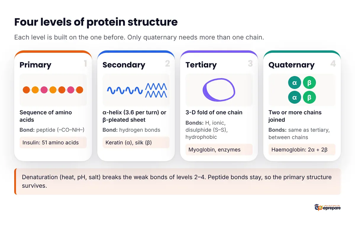
1. Primary structure: the sequence
The primary structure is the exact sequence of amino acids in a polypeptide chain. It is set by the DNA of the gene and read through mRNA. The only bonds involved are the peptide bonds between amino acids.
- Example: insulin has 51 amino acids in two chains: chain A with 21 and chain B with 30, joined by disulphide bridges.
- The primary structure decides every higher level. Change one amino acid and the whole shape can change.
- Sickle cell anaemia is the classic proof: in the β chain of haemoglobin, glutamic acid is replaced by valine at position 6. This lowers the haemoglobin's solubility, it forms fibres, and red blood cells are distorted into sickles.
2. Secondary structure: α-helix and β-pleated sheet
The polypeptide chain coils or folds into a regular, repeating pattern. These shapes are held mainly by hydrogen bonds between the C=O group of one peptide bond and the N–H group of another along the backbone.
- α-helix: the chain coils like a spring. There are about 3.6 amino acids per turn, and hydrogen bonds form between amino acids within the same coil. The keratin of hair is rich in α-helices.
- β-pleated sheet: lengths of the chain lie side by side in a zigzag (pleated) pattern, held by hydrogen bonds between the neighbouring strands. Silk protein (fibroin) is the standard example.
Many proteins contain both: some parts are helices, some are sheets, and the rest are loops joining them.
3. Tertiary structure: the 3-D fold of one chain
The helices and sheets of a single chain fold further into a specific three-dimensional shape. This fold is held by interactions between the R groups:
| Bond or interaction | Formed between | Strength and note |
|---|---|---|
| Disulphide bridge (–S–S–) | The sulphur-containing R groups of two cysteine amino acids | Covalent, so the strongest; not broken by mild heat |
| Ionic bond | Positively and negatively charged R groups (basic and acidic) | Broken by changes in pH |
| Hydrogen bond | Polar R groups | Weak individually, strong in large numbers; broken by heat |
| Hydrophobic interaction | Non-polar R groups | Non-polar groups are pushed together away from water, so they fold inward |
In a globular protein that sits in water, the non-polar R groups end up inside and the polar R groups stay on the surface facing water. That is why globular proteins are soluble. Myoglobin, the oxygen-binding protein of muscle, is a single chain with a heme group, so its highest level is tertiary.
Tertiary structure is also what makes an enzyme work. The active site is made of a small number of amino acids (about 3 to 12) that may be far apart in the sequence but are brought together by folding.
4. Quaternary structure: more than one chain
Quaternary structure exists only in proteins made of two or more polypeptide chains. The chains are held together by the same kinds of bonds as tertiary structure.
- Haemoglobin is the standard example: 4 polypeptide chains, 2 α and 2 β, each with its own iron-containing heme group, about 574 amino acids in total.
- Its four subunits allow cooperative oxygen binding, which a single chain like myoglobin cannot do.
- A protein with only one chain (myoglobin) has no quaternary structure.
| Feature | Haemoglobin | Myoglobin |
|---|---|---|
| Found in | Red blood cells | Skeletal and cardiac muscle |
| Job | Carries O2 to tissues and some CO2 back | Binds oxygen in muscle |
| Chains | 4 (2 α, 2 β), each with heme | 1, with heme |
| Highest level | Quaternary | Tertiary |
All four levels in one table
| Level | What it is | Main bonds | Example |
|---|---|---|---|
| Primary | Sequence of amino acids | Peptide bonds | Insulin (51 amino acids) |
| Secondary | α-helix or β-pleated sheet | Hydrogen bonds (backbone) | Keratin (α-helix), silk (β-sheet) |
| Tertiary | 3-D fold of one chain | Hydrogen, ionic, disulphide bonds, hydrophobic interactions | Myoglobin, enzymes |
| Quaternary | Two or more chains together | Same as tertiary, between chains | Haemoglobin (2 α + 2 β) |
Remember this: ePrepare's memory trick: Primary = Pearls on a string; Secondary = Spring or Sheet; Tertiary = a Tangled ball; Quaternary = a Quartet of chains (haemoglobin has 4).
Denaturation: when the shape is lost
Denaturation is the loss of a protein's specific 3-D shape. Heat, a change in pH, or salts break the weak bonds (especially hydrogen bonds, and ionic bonds with pH) that hold the secondary, tertiary and quaternary structure. The chain unfolds and the protein stops working.
- The peptide bonds are not broken, so the primary structure (sequence) remains. Denaturation is not digestion.
- A familiar example is egg white (albumin) turning solid and white when it is cooked.
- For enzymes, heat above the optimum temperature denatures the enzyme, the active site loses its shape, and the reaction rate falls sharply. In the cold an enzyme is only inactive, not denatured.
Fibrous vs globular proteins
Based on their overall shape, proteins are grouped into two types:
| Feature | Fibrous proteins | Globular proteins |
|---|---|---|
| Shape | Long strands or sheets | Compact, rounded |
| Solubility in water | Insoluble | Usually soluble |
| Role | Structural; strong and elastic | Metabolic and regulatory |
| Examples | Keratin, collagen, myosin, actin, fibrin, silk | Enzymes, insulin, antibodies, haemoglobin, myoglobin, albumin |
Trick from ePrepare's notes: Fibrous = Fibres and Framework (structure). Globular = Globe-shaped, Gets work done (enzymes, hormones, antibodies).
How MDCAT asks about protein structure
- Bond-to-level matching: "α-helix is stabilised by…" (hydrogen bonds). "Which bond is covalent and found in tertiary structure?" (disulphide).
- Which protein has quaternary structure: haemoglobin, not myoglobin.
- Number facts: 3.6 amino acids per turn of the α-helix; insulin 51 amino acids; haemoglobin 4 chains.
- Denaturation logic: which level survives heat? (primary).
- Sickle cell: which amino acid replaces which, at what position, and which level is first changed? (primary; glutamic acid → valine at position 6 of the β chain).
- Classify the protein: keratin and collagen as fibrous; enzymes and haemoglobin as globular.
Common mistakes
- "Secondary structure is held by peptide bonds." Wrong. Peptide bonds make the primary chain; α-helix and β-sheet are held mainly by hydrogen bonds.
- "The peptide bond is a C–O bond." Wrong. It is a C–N bond (–CO–NH–). C–O is the glycosidic bond of carbohydrates.
- "Myoglobin has quaternary structure like haemoglobin." Wrong. Myoglobin is one chain, so it is tertiary only.
- "Haemoglobin has four identical chains." Wrong. It has 2 α and 2 β chains.
- "In sickle cell, valine is replaced by glutamic acid." Wrong way round. Glutamic acid is replaced by valine.
- "Denaturation breaks peptide bonds." Wrong. It breaks the weak bonds holding the shape; the sequence stays.
- "Disulphide bonds form between any two amino acids." Wrong. They form between the sulphur-containing R groups of cysteines.
MDCAT-style practice MCQs
1. The α-helix and β-pleated sheet of a protein are held mainly by:
A. Peptide bonds
B. Hydrogen bonds
C. Disulphide bonds
D. Ionic bonds
Show answer
B. Secondary structure is stabilised by hydrogen bonds between C=O and N–H groups of the backbone.
2. Which protein has quaternary structure?
A. Myoglobin
B. Haemoglobin
C. Keratin strand
D. A single polypeptide enzyme
Show answer
B. Haemoglobin has four chains (2 α, 2 β); myoglobin is a single chain with tertiary structure only.
3. The primary structure of a protein is:
A. Its 3-D shape
B. The coiling of the chain into a helix
C. The sequence of amino acids
D. The number of chains it has
Show answer
C. Primary structure is the exact amino acid sequence, held by peptide bonds and set by the gene.
4. The number of amino acids per turn of an α-helix is about:
A. 2.6
B. 3.6
C. 4.6
D. 5.4
Show answer
B. An α-helix has about 3.6 amino acids in each turn.
5. A covalent bond that helps hold the tertiary structure of a protein is the:
A. Hydrogen bond
B. Ionic bond
C. Disulphide bridge
D. Hydrophobic interaction
Show answer
C. The disulphide bridge (–S–S–) between two cysteines is covalent; the others are weak, non-covalent interactions.
6. When a protein is denatured by heat, which level of structure remains?
A. Primary
B. Secondary
C. Tertiary
D. Quaternary
Show answer
A. Heat breaks hydrogen and other weak bonds, but the peptide bonds and the sequence remain.
7. In sickle cell anaemia, the change in the β chain of haemoglobin is:
A. Valine replaced by glutamic acid at position 6
B. Glutamic acid replaced by valine at position 6
C. Glycine replaced by valine at position 6
D. Loss of the heme group
Show answer
B. One amino acid change in the primary structure (glutamic acid → valine) alters the shape and behaviour of haemoglobin.
8. Which group contains only fibrous proteins?
A. Keratin, collagen, myosin
B. Insulin, antibodies, haemoglobin
C. Enzymes, albumin, myoglobin
D. Collagen, insulin, haemoglobin
Show answer
A. Keratin, collagen and myosin are insoluble structural (fibrous) proteins; the others contain globular proteins.
Learn it in ePrepare's conceptual notes
The proteins lesson in the Biological Molecules chapter of ePrepare's Biology conceptual notes covers amino acid structure, the peptide bond (with a drawn dipeptide), a four-panel diagram of the levels of structure, fibrous and globular proteins, the roles of proteins, haemoglobin vs myoglobin and sickle cell anaemia. Each section has easy-Urdu explanations, memory tricks, common mistakes and practice MCQs with explanations.
See how the notes are organised in ePrepare's MDCAT conceptual notes, and where Biological Molecules sits among the chapters that carry the most marks in MDCAT Biology high-yield chapters. Protein shape links straight to enzyme action and feedback inhibition, covered in negative feedback in hormonal control, and to genetics in sex-linked inheritance.
Quick revision table
| Point | Remember |
|---|---|
| Monomer | About 20 amino acids; H2N–CH(R)–COOH |
| Peptide bond | –CO–NH– (C–N); formed by condensation, water released |
| Primary | Sequence; peptide bonds; insulin 51 (21 + 30) |
| Secondary | α-helix (3.6 per turn) and β-sheet; hydrogen bonds |
| Tertiary | 3-D fold of one chain; H, ionic, disulphide, hydrophobic |
| Quaternary | 2 or more chains; haemoglobin 2 α + 2 β |
| Myoglobin | One chain; tertiary only |
| Denaturation | Heat, pH, salt break weak bonds; primary stays |
| Fibrous / globular | Insoluble, structural / soluble, metabolic |
| Sickle cell | Glutamic acid → valine at position 6 of the β chain |
آسان الفاظ میں: primary structure صرف amino acids کی ترتیب ہے، جو peptide bonds سے جڑی ہوتی ہے۔ Secondary میں یہ chain سپرنگ کی طرح α-helix بناتی ہے یا β-sheet کی طرح تہہ ہو جاتی ہے، اور یہ hydrogen bonds سے قائم رہتی ہے۔ Tertiary میں پوری chain ایک 3-D شکل میں مڑ جاتی ہے۔ Quaternary تب ہوتی ہے جب ایک سے زیادہ chains مل کر ایک protein بنائیں، جیسے haemoglobin کی چار chains۔ گرمی سے protein کی شکل بگڑ جاتی ہے (denaturation)، لیکن amino acids کی ترتیب وہی رہتی ہے۔
Frequently asked questions
What are the four levels of protein structure?
Primary (the amino acid sequence), secondary (α-helix and β-pleated sheet), tertiary (the 3-D folding of one chain) and quaternary (two or more chains joined, as in haemoglobin).
Which bonds are found in each level?
Primary: peptide bonds. Secondary: hydrogen bonds. Tertiary and quaternary: hydrogen bonds, ionic bonds, disulphide bridges and hydrophobic interactions between R groups.
Does every protein have quaternary structure?
No. Only proteins made of two or more polypeptide chains have it. Myoglobin is a single chain, so its highest level is tertiary.
What happens to protein structure during denaturation?
The weak bonds that hold the secondary, tertiary and quaternary levels break, so the protein unfolds and loses its function. The peptide bonds, and therefore the primary structure, are not broken.
What is the difference between fibrous and globular proteins?
Fibrous proteins are long, insoluble and structural, like keratin and collagen. Globular proteins are compact, usually soluble and do metabolic work, like enzymes, antibodies and haemoglobin.
Which chapter is protein structure in for MDCAT?
It is in the Biological Molecules chapter of Biology, and it links to Enzymes. You can check the full topic list in the MDCAT syllabus guide.